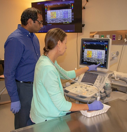
Sound waves may help diagnose wooden breasts in chicken
Collaborative veterinary/poultry science research leads to exploring sound in data interpretation.
November 29, 2018

Auburn University faculty collaborating on a joint veterinary medicine/poultry science research project have discovered the music in their science, which could help visually impaired scientists interpret image data, according to an announcement from the Alabama university.
Veterinarian Rachel Moon, an assistant clinical professor of radiology in the Auburn College of Veterinary Medicine, and Amit Morey, an assistant professor in the department of poultry science in the College of Agriculture, found an unexpected linkage between science and music while developing novel detection methods of their laboratory work.
They are studying an economically undesirable muscular condition in broiler chickens known as muscle myopathy, commonly referred to as wooden or woody breast.
“Woody breast is a condition that results in tough, chewy breast meat in the broiler chicken,” Morey said. “The condition is undesirable to the consumer, and it currently affects more than 10% of the fast-growing broiler segment of the poultry industry.
“In the U.S., we are producing some 9 billion broilers every year. These birds are more efficient and faster growing, which has resulted in occurrence of novel muscle myopathies such as the woody breast. We are looking for ways to quickly and accurately detect the condition so that the meat can be removed from the market before it causes issues with consumer rejection,” he said.
Morey, who specializes in the development of novel detection methods in poultry science, said he learned of technology available at the Auburn College of Veterinary Medicine that could help and reached out to Moon, a board-certified veterinary radiologist.
The team is using Moon’s ultrasound technology specialty as a detection means. “We are using ultrasound and a more advanced process called elastography as a means of generating images of the breast meat,” Moon said.
The technology, typically used to identify medical conditions in animals, produces vivid image graphs of the chicken breast that depict varying differences in the muscular structure between normal and muscles affected by woody breast, the researchers explained.
While the project has produced successful results for that particular industry problem, it also had a somewhat unexpected twist: the creation of music from the research.
Morey has been working as a mentor to Johns Creek, Ga., high school student Divya Srinivansan, who has an interest in converting visual imagery into auditory frequencies that can be used by visually impaired researchers and diagnosticians. The high school student’s work has won her several awards at national and international competitions, including the 2018 Intel International Science & Engineering Fair, Auburn said.
After evaluating various online free software to convert images into sound, Srinivansan chose Photosounder to convert the ultrasound images into frequency profile graphs and sound.
“The sounds from the software were very harsh, and I wanted to convert the graphs to actual musical notes,” Morey said.
Lakshmi Krishnaprasad, a former computer science and music student at Auburn, translated the frequency profiles of ultrasound images to musical tones.
“Krishnaprasad derived an abstract interpretation from the graphs to musically express the general curves of the ultrasound graphs,” Morey said.
For the ultrasound images showing a normal chicken breast structure, the C major arpeggio — containing the notes C, E and G — was used to represent the initial ascension of the graph, followed by an immediate jump to a high pitch and then dropping back to the low octave. For the ultrasound image showing the structure of severe woody breast muscle, an instrumental concept was used called overtones, where multiple notes can be played using only one fingering, the researchers explained.
“It really added an unexpected dimension to our research,” Morey said.
Moon explained that an ultrasound image is actually derived from auditory waves, but these are at very high frequencies that the human ear cannot detect.
“The prospect of turning ultrasound images into usable auditory data is exciting,” Moon said. “Ultrasound images are based on very-high-frequency sound waves that the human ear is incapable of hearing. With the aid of our two student co-investigators, we are taking that scientific visual data and turning it back into sound waves that are within the auditory range of the human ear. That is a very intriguing aspect of this project.”
You May Also Like


