Method may provide a good model for studying how similar viruses swap genetic material through reassortment.
April 28, 2020

Live cell imaging has been used by researchers at The Pirbright Institute in the U.K. to reveal new information about how different strains of infectious bursal disease virus (IBDV) might swap genetic material, a process that could lead to new strains with the potential for causing more severe disease, the institute said in a recent announcement.
IBDV is a type of birnavirus that causes an economically important disease of poultry, known as Gumboro disease. It infects both domestic and wild birds, and targets the immune system tissues, weakening the resistance of chickens to infection by other microbes, Pirbright explained.
The virus genome is split into two segments of double stranded RNA, a molecular relative of DNA. During replication, new copies of the IBDV genome are made in discreet sites within cells called "virus factories" (VFs), Pirbright said.
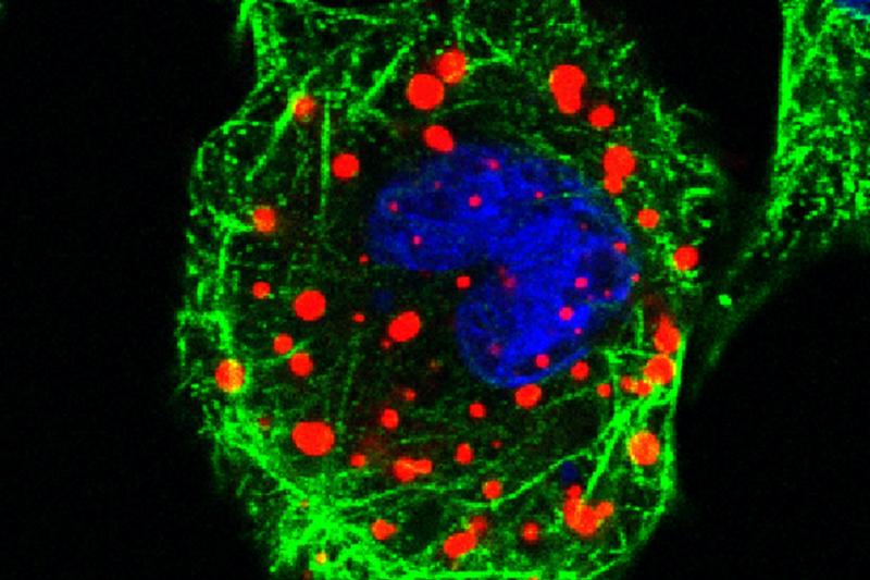
In order to further study IBDV replication, Pirbright scientists created the first genetically engineered IBDV viruses that emit red or green fluorescent light so that they could track the location and movements of the VFs using live cell imaging microscopy.
Their experiments, the results of which have been published in the Journal of Virology, revealed that the VFs are very dynamic and move around within the cytoplasm of an infected cell. Over time, these structures merge and combine with each other, leading to fewer, but larger, VFs, Pirbright noted.
VFs have been thought to represent a barrier to genome mixing between two different strains infecting the same cell, a process known as reassortment. When the team at Pirbright co-infected cells with red and green tagged viruses, they found that while the red and the green viruses started off in separate VFs, they eventually merge so that red and green viruses were located in the same VFs, the institute reported.
They also found that the merging of VFs occurs at least 10 hours after the cells had been infected, which suggests that the potential for IBDV to swap the two segments of its genome occurs late in the viral replication cycle.
“Our study sheds new light on the replication of IBDV, and possibly other birnaviruses, revealing that the VFs are highly dynamic structures that interact with one another, which may provide the opportunity for different strains of IBDV to swap segments of their genome. The next step is to understand more about the molecular factors that are involved in and control this process,” Dr Andrew Broadbent, head of the birnavirus group at Pirbright, said.
The methods and observations in this study may also provide a good model for studying how viruses with similar genome structures, such as bluetongue virus and African horse sickness virus, swap genetic material through reassortment.
You May Also Like


.png?width=300&auto=webp&quality=80&disable=upscale)
